Tibialis posterior muscle exercises. Tibialis posterior muscle Tibialis posterior muscle how to train a child
Karina Grishanova 7.05.2015 2597

If you dream of a beautiful walk, these exercises are necessary for you.
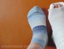
It's time for open sandals and high heels. To feel confident in such shoes, it is necessary to strengthen the muscles of the foot and ankle joint. Regular exercise will make your gait stable and beautiful, and will also protect you from injuries that can occur when walking or running.
What muscles need to be trained?
There are 5 main muscles involved in the movement of the foot:
- gastrocnemius And soleus provide flexion of the foot on the side of the sole;
- anterior tibial unbends the foot;
- peroneus muscle not only bends the foot, but also takes it to the side;
- posterior tibialis muscle responsible for stabilizing the ankle joint.
To walk beautifully and confidently, you need to work on all these muscles.
Exercise examples
We bring to your attention several exercises aimed at strengthening the muscles of the foot. Their main advantage is that they can be performed at home or in the office: no special devices are required for this.
Stop extension
Initial position: sitting on the floor, straight legs extended in front of you. Pull your feet alternately away from you and towards you. Make sure that during the exercise, the heel, thumb and little finger remain in the same plane. Do not curl your toes under the arch of your foot. Repeat 10 times.
Circular foot movements
Initial position: former. Fulfill circular motions feet first inward, then outward. Try to touch the floor with a bone thumb when rotating inward and with the little finger when rotating outward. Do 10 movements in each direction.
knee exercise
Initial position: kneeling on the floor. From the starting position, sit on your feet so that the bones of the big toes and the heels of both feet are close to each other. Stay in this position for 1 minute.
Towel exercise
Initial position: Lay out a medium-sized rectangular towel on the floor and stand on one of its ends. Keeping your heels on the floor, gradually pull the towel towards you with your toes. Then straighten the towel and repeat the exercise 10 times.
Finger lift
Initial position: standing, back straight. Raise up on your toes as high as you can. Keep your heels in the air and begin to gradually bend your knees. In a semi-squat position, place your heels on the floor and only then straighten your legs. The knees and ankles should remain in a straight position, without deviations outward or inward. Repeat 10 times.
If you can’t do the exercises regularly, practice picking up small objects from the floor with your toes as often as possible. You can do this even at work: scatter pencils or paper clips under the table and study.
How often to exercise?
For the most effective exercises, you need to perform them every day. Do not worry: the whole complex will take you no more than 10 minutes, but your gait will improve in a couple of weeks.
The tibialis posterior muscle (TMP) is located between the fibula and tibia and is attached to the interosseous septa. She occupies the deepest position. Above it are the flexor muscles of the fingers, the flexor of the thumb and the muscles of the lower leg. Its base is closer to the heads of the tibia.
MBA functions:
- Formation of the arch of the foot, lifting it up. This is an isolated movement.
- Stabilization of the fibula. If the fibula is not fixed to the right degree, it will wobble. Friction between the head of the fibula and tibia causes instability of the knee. Gradually, this leads to arthrosis of the knee joint. There is also instability in the ankle joint, the position of the talus changes. It shifts slightly forward, which limits the flexion and extension of the foot. This is especially important for athletes, as there is a shortening of the stride during walking and running. If this phenomenon is observed in one leg, over time it leads to the formation of a difference in the volume and tone of the buttocks.
- Holds the arch of the foot and stabilizes the knee joint.
It may seem that the MBA is just holding the vault. But if its functions are weakened, the location of the thigh, knee, bones of the lower leg are changed. This leads to various pathological changes in the skeleton and pain, impaired posture, degenerative changes in the spine.
With weakness of the MBA, other muscles cannot work correctly: neither the gluteus nor the extensors of the fingers. They turn off during movement, during a step. This causes pain and discomfort, ultimately leading to a decrease in the mobility of the lower leg.
Weakness of the posterior tibial muscle provokes weakness of the ligamentous apparatus of the foot, all the small bones that form its arch diverge to the sides, this leads to flat feet.
The transverse and longitudinal arch of the foot forms the tone of this muscle. The efficiency of all legs depends on it.
MBA training principles
To restore the functions of the MBA, you need to perform a special set of exercises to reduce it, as well as bring it into tone with each step. This is possible if the foot is well extended with each step.
The isolated movement of the MBA occurs when the foot moves to the side inward. This is how only the tibialis posterior muscle works.
For athletes and those who have weakened muscle tone, if flat feet, it is important to follow the principles of training and exercise regularly, this is the only way to achieve results.
How to restore the tone of the posterior tibial muscle
It is very difficult to influence the MBA from the outside. It can be approached between the calf muscle and the tibia.
Massage can be done independently by doing the following:
- tapping on the posterior tibia from bottom to top and vice versa. At the same time, you need to move your foot to the left and right. This will enhance the effect. Tapping should be gentle, the use of force will only cause harm;
- pressure with the thumbs or the base of the palm along the tibia with inside from the base of the MBA to the foot. Closer to the ankle joint in this place are nerve endings and pressure can be very painful.
It is necessary to work out the muscle well with a massage, relax tense areas and then proceed to the exercises.
For the treatment of flat feet, it is necessary to start classes on maintaining and restoring the tone of the MBA with static exercises.
- Sitting put your feet on the floor. On the foot from the inside in the toe area you need to press. The foot at this time exert resistance for a few seconds. In this case, the knee does not move, it remains in a static position. Perform up to 10 repetitions.
- Sitting on a chair, make movements with the foot, without lifting it from the floor, inward to the other foot. The heels are in place, the toes move towards the opposite leg. Do 10 repetitions for each leg.
- Do exercise 2, but at the same time with two feet. When fingers touch, press against each other for 3 seconds. The feet seem to be trying to move inward, but interfere with each other. Perform 5 to 10 repetitions.
The second stage of training - exercises for the posterior tibial muscle with rubber band.
- Fasten the tourniquet and make a loop. Put the loop on the foot, while sitting on the floor, the leg is extended forward. Make an isolated movement with the foot (rotation of the socks inward with effort) with the tourniquet thrown over. The number of times individually, until fatigue in the muscle. This version of the exercise can be performed while sitting on a chair.
- This movement must be connected when the first exercises after the fracture are performed with ease. You need to put your foot on the step along the edge. It is desirable that half of the foot hangs slightly from the dais. Now you need to rise a little, shifting the body weight to the foot of the working leg, then return to the starting position. Perform to fatigue, it is important not to overdo it.
To restore and maintain the tone of the MBA, you need to carefully and calmly perform the entire complex and massage every day. Regular training of the tibial muscle will return it to its former functionality.
Manufacturer of refrigeration units. Xiron-Holod buy a chiller www.xiron.ruAnatomy of the lower leg
The shin is part lower limb and is located between the knee and foot. The lower leg is formed by two bones - the tibia and the fibula, which are surrounded by muscles on three sides that move the foot and fingers.
Lower leg bones
Tibia
The tibia at its upper end expands, forming the medial and lateral condyles. On the top of the condyles are the articular surfaces that serve for articulation with the condyles of the thigh; between them is the intercondylar eminence. Outside, on the lateral condyle there is an articular surface for articulation with the head of the fibula. The body of the tibia is like a trihedral prism, the base of which is turned backwards; it has three surfaces corresponding to the three sides of the prism: inner, outer and back. Between the inner and outer surfaces is a sharp leading edge. In its upper section, it passes into a well-defined tibial tuberosity, which serves to attach the tendon of the quadriceps femoris. On the back surface of the bone is a rough line of the soleus muscle. The lower end of the tibia expands and on the inside has a protrusion directed downwards - the medial malleolus. On the distal epiphysis of the tibia is the lower articular surface, which serves for articulation with the talus.
Fibula
The fibula is long, thin and located laterally. At the upper end, it has a thickening, the head, which articulates with the tibia, at the lower end it also has a thickening, the lateral malleolus. Both the head and the malleolus of the fibula protrude outward and are easily palpable under the skin.
Joints of the bones of the lower leg
Between both bones of the lower leg - the tibia and fibula - is the interosseous membrane of the lower leg. The head of the fibula is articulated with the tibia by means of a joint that has a flat shape and is reinforced in front and behind by a ligamentous apparatus. The lower ends of the leg bones are connected by syndesmosis. The joints between the bones are inactive.
Leg muscles
On the lower leg, the muscles are located on three sides, making up the anterior, posterior and outer groups. The anterior muscle group extends the foot and fingers, and also supinates and adducts the foot. These include the tibialis anterior, extensor digitorum longus, and extensor hallucis longus. The posterior muscle group that flexes the foot and toes is: triceps calf, flexor digitorum longus and flexor hallucis longus, tibialis posterior, hamstrings. The outer group of muscles abducts, pronates and flexes the foot; it includes the long and short peroneal muscles.
Tibialis anterior
The tibialis anterior muscle originates from the outer surface of the tibia, the interosseous membrane, and the fascia of the leg. Going down, it passes under two ligaments located in the area of the ankles and ankle joint - the upper and lower retainers of the extensor tendons, which are places of thickening of the fascia of the lower leg and foot. Attaches the anterior tibial muscle to the medial sphenoid bone and the base of the first metatarsal bone. This muscle is well felt under the skin throughout, especially in the transition area from the lower leg to the foot. Here, her tendon protrudes when the foot is extended. The function of the anterior tibial muscle is that it contributes not only to the extension of the foot, but also to its supination.
Long finger extensor
The extensor digitorum longus lies outside the tibialis anterior in the upper leg. It starts from the upper end of the tibia, the head and anterior edge of the fibula, as well as from the interosseous membrane and fascia of the leg. Passing to the foot, this muscle is divided into five tendons, of which four are attached to the distal phalanges of the second, third, fourth and fifth fingers, and the fifth - to the base of the fifth metatarsal bone.
Function extensor longus fingers as a multi-joint muscle consists not only in the extension of the fingers, but also in the extension of the foot. Due to the fact that one of the tendons of the muscle is attached to the outer edge of the foot, it not only unbends, but also somewhat penetrates the foot.
Long extensor thumb
The long extensor of the thumb starts from the inner surface of the fibula and the interosseous membrane in the region of the lower half of the leg. This muscle is weaker than the two previous ones, between which it is located. It is attached to the base of the distal phalanx of the thumb. The function of the muscle is that it is an extensor not only of the big toe, but of the entire foot, and also contributes to its supination.
Triceps muscle of the leg
The triceps muscle of the leg is located on the back of the leg and has three heads. Two of them make up the superficial part of this muscle and are called calf muscle, and the deep one forms the soleus muscle. All three heads pass into one common, calcaneal (Achilles) tendon, which is attached to the tuber of the calcaneus.
Starting point calf muscle are the medial and lateral condyles of the femur. Its medial head is better developed and descends somewhat lower than the lateral one. The function of these heads is twofold: flexion of the lower leg at the knee joint and flexion of the foot at the ankle joint.
The soleus muscle originates from the posterior surface of the upper third of the body of the tibia, as well as from the tendon arch located between the tibia and fibula. This muscle is located deeper and somewhat lower than the calf muscle. Passing behind the ankle and subtalar joints, the soleus muscle causes flexion of the foot.
The triceps muscle of the lower leg is clearly visible under the skin and is easily palpated. The calcaneal tendon protrudes significantly posterior to the transverse axis of the ankle joint, due to which the triceps muscle of the leg has a large moment of rotation in relation to this axis.
The medial and lateral heads of the gastrocnemius muscle are involved in the formation of the popliteal fossa, which has the shape of a rhombus. Its boundaries are: above and outside - biceps thighs, above and inside - a semimembranosus muscle, and below - two heads of the gastrocnemius muscle and plantar muscle. The bottom of the fossa is the femur and the capsule of the knee joint. Through the popliteal fossa pass the nerves and blood vessels that feed the lower leg and foot.
Long finger flexor
The long flexor of the fingers starts from the posterior surface of the tibia and passes to the foot under the medial malleolus in a special channel located under the ligament - the retainer of the flexor tendons. On the plantar surface of the foot, this muscle crosses the tendon long flexor thumb and after joining it square muscle the sole is divided into four tendons, attached to the bases of the distal phalanges of the second to fifth fingers.
The function of the long toe flexor is to flex and supinate the foot and to flex the toes. It should be noted that the square muscle of the sole, attached to the tendon of this muscle, contributes to the "averaging" of its action. The fact is that the long flexor of the fingers, passing under the medial malleolus and fan-shaped dividing towards the phalanges of the fingers, causes not only their flexion, but also some reduction to the median plane of the body. Due to the fact that the square muscle of the sole pulls the tendon of the long flexor of the fingers outward, this adduction is somewhat reduced and the flexion of the fingers occurs to a greater extent in the sagittal plane.
flexor thumb longus
The long flexor of the thumb is the most strong muscle among all the deep muscles of the back of the leg. It starts from the lower part of the posterior surface of the fibula and the posterior intermuscular septum. On the plantar surface of the foot, this muscle is located between the heads short flexor thumb. It is attached to the plantar surface of the base of the distal phalanx of the thumb.
The function of the muscle is to flex the thumb and the entire foot. Due to the fact that the tendon of the muscle partially passes into the tendon of the long flexor of the fingers, it has some effect on the flexion of the second and third fingers. An increase in the moment of rotation of the long flexor of the thumb is facilitated by the presence of two large sesamoid bones on the plantar surface of the metatarsophalangeal joint of the thumb.
Tibialis posterior
The tibialis posterior muscle is located under the triceps muscle of the lower leg. It starts from the posterior surface of the interosseous membrane of the lower leg and adjacent areas of the tibia and fibula. Passing under the medial malleolus, this muscle attaches to the tuberosity of the navicular bone, to all the cuneiform bones and to the bases of the metatarsal bones. Its function is to flex the foot, adduct and supinate it.
Between the posterior tibial and soleus muscles is the shin-popliteal canal, which looks like a gap and serves to pass blood vessels and nerves.
Hamstring
The popliteal muscle is a short flat muscle directly adjacent to the back of the knee joint. It starts from the lateral condyle of the thigh, below the gastrocnemius muscle, and the bag of the knee joint, goes down and inward and attaches to the tibia above the line of the soleus muscle. The function of this muscle is that it contributes not only to flexion of the lower leg, but also to its pronation. Due to the fact that this muscle is partially attached to the capsule of the knee joint, it pulls it back as the lower leg flexes.
Peroneus longus muscle
The long peroneal muscle has a pinnate structure. It lies on the outer surface of the fibula, starts from its head, partly from the fascia of the leg, from the lateral condyle of the tibia and from the outer surface of the fibula in the region of its upper two thirds. In the lower third, the muscle covers the short peroneal muscle. The tendon of the long peroneal muscle wraps around the back and bottom of the lateral malleolus. In the region of the outer surface of the calcaneus, the muscle is held by ligaments - the upper and lower retainers of the tendons of the peroneal muscles. Passing to the plantar surface of the foot, the tendon of the muscle goes along the groove located on the lower surface of the cuboid bone, and reaches the inner edge of the foot. The peroneus longus attaches to the tuberosity on the undersurface of the base of the first metatarsal, to the medial cuneiform and to the base of the second metatarsal.
The function of the muscle is to flex, pronate, and abduct the foot.
Peroneus brevis
The short peroneal muscle originates from the outer surface of the fibula and intermuscular septa shins. The tendon of the muscle bends around the lateral malleolus of the lower leg from below and behind and is attached to the tuberosity of the fifth metatarsal bone. The function of the short peroneal muscle is to flex, pronate, and abduct the foot.
References
- human anatomy: studies. for stud. inst. physical cult. / Ed. Kozlova V.I. - M., "Physical culture and sport", 1978
- Sapin M.R., Nikityuk D.K. Pocket atlas of human anatomy. M., Elista: APP "Dzhangar", 1999
- Sinelnikov R. D. Atlas of human anatomy: in 3 volumes. 3rd ed. M.: "Medicine", 1967
This syndrome is the cause of medial hindfoot pathology, often overlooked and misdiagnosed, especially in the early stages. This is a direct result of the loss of function of the tibialis posterior tendon.
Chronic inflammation leads to degeneration and stretching of the tendon with interstitial edema, thinning, and chronic injury to the tendon. If left untreated, all this leads to a violation of the alignment of the hindfoot and midfoot with heel pronation, plantaflexion of the talus, subluxation in the talonavicular joint and, as a result, the formation of a unilateral flat foot.
The tibialis posterior muscle is active during the stance phase, fires immediately after heel-to-foot contact, and quickly stops contracting after heel lift. Its abdomen begins deep inside the posterior part of the lower limb, the tendon runs down to the posterior part of the medial malleolus, where it lies anterior to the flexor digitorum longus tendon, the posterior tibial neurovascular bundle (posterior posterior artery, vein, and nerve), and the flexor hallucis tendon. All of these structures are limited to the flexor retinaculum near the medial malleolus. The tendon of the posterior b/b muscle runs in a groove behind and below the medial malleolus, dividing into 3 parts at the medial side of the talus. The anterior part is attached to the tuberosity of the scaphoid, the middle part continues into the plantar tarsal region and is attached to the plantar part of the sphenoid bones, the cuboid and at the base of the 2,3 and 4 metatarsals. Rear end is introduced as a bundle into the anterior part of the inferior calcaneal-navicular ligament. The medial malleolus acts like a multi-roller unit, allowing the posterior calf tendon to change direction of pull, and these attachment points provide supination for the hindfoot and midfoot during weight transfer while stabilizing the midfoot arch structure.
The main function of the posterior b/b muscle is to achieve supination in the subtalar joint and adduction of the forefoot around the oblique axis of the midmetatarsal joint.
. At the beginning of the stance phase, the posterior ankle muscle contracts eccentrically to slow the pronation that occurs at the subtalar joint and during internal rotation of the ankle bone.
. During the mid-stance period, the muscle contracts concentrically, providing stability to the mid-metatarsal joint in preparation for the push.
. On heel lift, this provides plantar torque that allows the heel to lift off the ground.
Thus, the posterior pelvic muscle acts as a primary stabilizer against posterior valgus, anterior abduction, and as an antagonist to the peroneal muscles, especially the peroneus brevis.
Etiology.
The reasons are unclear, associated with the following conditions:
. Obesity
. Excessive pronation of the foot, which tends to compress and disrupt the blood supply to the tendon that wraps around the medial malleolus deep under the support.
. Structural and anatomical abnormalities, eg, accessory navicular bone, rigid or mobile flatfoot, osteophyte proliferation in the medial malleolus sulcus, shallow sulcus, and ankle equinus.
. Inflammatory diseases joints, RA, seronegative arthritis
. Collagen vascular diseases
. Direct trauma where the tendon is torn by fragments of the medial malleolus
. Indirect trauma such as ankle fracture, eversion ankle sprain, acute avulsion injury to the navicular, and posterior patellar tendon dislocation
. Iatrogenic effect (injection of steroids into the area)
Pathology.
The idea of dysfunction can be divided into 4 stages:
1. Asymptomatic stage. Evaluation of the patient may reveal underlying disorders that may lead to the development of dysfunction. For example, fully compensated varus of the hindfoot, or obesity.
2. Stage initial symptoms. Tendonitis (inflammation of the tendon sheath in the area of the flexor retinaculum). Mild muscle weakness.
3. Stage of severe dysfunction. It is characterized by damage inside the tendon, elongation without damage, even separation of the tendon from the boat. Pronounced pronation of the middle section and abduction of the anterior section.
Other classification:
. acute phase. It lasts 2 weeks after the onset, during which tendon pathology may not be diagnosed. Typical: diffuse edema, softness on the medial side of the ankle joint. There may be soreness and fatigue of the muscles of the lower limb.
. Subacute phase. Lasts from 2 weeks to 6 months. There is pain and swelling along the tendon, from the back of the medial malleolus to the medial longitudinal arch. It may also be a symptom of tarsal tunnel syndrome due to local nerve compression. Passive movements in the subtalar and midmetatarsal joints usually do not cause pain, but the gait changes, there is no push, the anterior section is retracted, and there is no supination when the heel and toes are torn off.
. chronic phase. Comes in about 6 months. Patients have a unilateral rigid flat foot. In an advanced case, the pain may move from the medial to the lateral part of the tarsal sinus. Lateral pain occurs due to progressive valgus deformity of the hindfoot, which leads to calcaneofibular axial loading, periosteal inflammation, peroneal tendonitis, and subtalar tendinitis.
clinical picture.
Approximately 50% of cases are preceded by local trauma - severe eversion of the posterior section.
Women over 40 and younger athletes are more likely to suffer.
Patients often do not seek help in the early stages, in 1 or in the acute phase, because. symptoms are mild.
. Patients usually present in stage 2 or subacute phase, with diffuse edema and heat in the medial ankle and along the tendon. Patients experience difficulty or a feeling of instability when the heel comes off on the affected side, the heel does not supinate when it comes off the surface.
. In stage 3 or in the chronic phase, the patient notices a gradual decrease in the height of the longitudinal arch, the development of a flat foot on one side, fatigue in the lower limb when walking. When viewed from behind, there is excessive abduction of the forefoot (too many toes symptom). In severe cases, loss of longitudinal arch, eversion of the calcaneus. Excessive wear on the medial part of the heel in shoes.
Diagnosis and differential diagnosis.
The integrity of the posterior pelvic tendon is assessed by palpation with the patient actively plantar flexing and adducting the foot and the clinician applying abductive force to the forefoot. It is important to determine the exact site of damage within the tendon and compare it with a healthy foot. Direct pressure along the course of the tendon may reveal pain, and active inversion of the foot against resistance may reveal decreased posterior valvular muscle strength. If there is a partial injury to the tendon, it can be palpated.
If the tendon is completely damaged, the tendon will not be palpable along its normal bed, and the patient cannot invert the foot against resistance.
Partial or complete damage due to trauma is accompanied by various pains in the tuberosity of the navicular bone. Overuse injury and tendon degeneration present with pain distal to the medial malleolus.
MRI is the most useful method for examining the tendons around the ankle and identifying injuries. Other diagnostic tests include bone scans and injection of a radiocontrast agent into the tendon sheath. Early diagnosis is not improved by a direct radiograph, however, a review of the foot will show the extent of structural changes in stage 3. A standard anteroposterior radiograph shows an increase in the angle between the longitudinal axis of the talus and the longitudinal axis of the calcaneus, abduction of the forefoot, and displacement of the 2nd metatarsal. The long axis of the forefoot no longer bisects the angle of the hindfoot. Normally, the linear relationship between the talus, navicular, medial sphenoid and first metatarsal is lost on the lateral radiograph.
If the situation progresses, then osteoarthritis of the 1st metatarsophalangeal joint, secondary to the hallux limitus, appears.
Diffdiagnosis should rule out:
bone anomalies
1. scaphoid syndrome (os tibiale externum), trihedral bone syndrome, scaphoid avulsion, scaphoid stress fracture
2. osteochondritis or avascular necrosis of the head of the ram or rook
3. fracture of the medial malleolus
4. subtalar tarsal coalition
5. inflammation of the medial sinus tarsi
soft tissue disorders:
1. deltoid ligament sprain
2. medial capsulitis of the ankle
3. tarsal tunnel syndrome
4.Thumb flexor longus or long finger flexor sprain
5. posterior calcaneal bursitis
Other cases of unilateral flat foot (true or relative leg length difference, tarsal coalition) should also be considered in the diagnosis.
Treatment.
Treatment depends on the stage or phase of the disease. Treatment must be carried out quickly and aggressively to prevent further deterioration. In the early stages - reducing inflammation, stabilizing the joint, controlling pain - up to 8 weeks. In more severe and persistent cases, it is possible surgical recovery tendons with fixation of the joint.
Conservative treatment: NSAID, ultrasound, hindfoot taping in inversion position to relieve tension in tendons. Orthotics with soft temporary orthoses (valgus pillows, medial full foot pillows - cobra) are used to invert the hindfoot. Individual rigid anti-pronator orthoses allow the posterior b/b muscle to function more efficiently, because. directed at the underlying pathomechanical defect. The brace controls movement in the subtalar joint by reducing tendon strain by controlling anterior abduction (using a lateral collar). Exercise therapy is aimed at strengthening the back b / w muscle. In more severe cases, immobilization of the foot in an inverted position in a cast up to the knee is required for several weeks. Steroid therapy is not used because of the potential for damage to an already weakened tendon.
Surgical treatment is indicated in the 2nd stage or in the subacute phase. In the absence of effect from 8 weeks of conservative therapy or in the 3rd stage and 4th phase. In stubborn cases with mild tenosynovitis but no apparent tendon injury, tendon release, synovectomy, is indicated.
Synovectomy, strengthening of the tendon insertion, or transfer of the flexor digitorum longus is indicated in more severe cases characterized by tendon lengthening.
Severe cases with complete injury or fibrosis of the tendon are treated with flexor digitorum longus grafting, shortening of the ligaments and talonavicular capsule, and surgical widening of the bony canal under the medial malleolus. Arthrodesis of the hindfoot joints, such as between the talus and calcaneus, subtalar fusion, talonavicular fusion, or combined talonavicular and calcaneonavicular fusion may be indicated in advanced stages with lateral hindfoot pain.
The results of corrective surgery are not always direct. The procedure requires a long period of recovery, rehabilitation, exercise. It is difficult to accurately predict the degree of post-operative correction of planovalgus in degrees, however, an increase in stability during the period of support can be expected. Osteoarthritis of the joints in the back of the foot develops after a long time, because. If normal joint alignment is disturbed due to arthrodesis, the repaired tendon may weaken in the future with a return of preoperative symptoms.
This is the main muscle that holds the arch of the foot (see Fig. 58). It is with her weakness that the development of flat feet is associated. In addition to the fact that she takes a major part in the formation of the arch of the foot, she also stabilizes the fibula, thereby taking part in the stabilization of the knee and ankle joints.
With weakness of the posterior tibial muscle (TBB), the arch of the foot drops, the stabilization of the ankle and knee joints is disturbed, which leads to injuries of these joints and ligaments of the joints, even with a slight load. Pain in the knee joint is very often associated with weakness of this muscle. She is also one of the first to get tired when walking for a long time, causing instability of the foot and knee joint.
The two most common causes of weakness in this muscle are a violation of innervation and injury to the muscle itself.
Very often you can now hear about the relationship of all parts of our body. Some of these connections have nothing to do with reality. Some are quite minor. But the third group really makes an impact. These connections include the foot.
The foot is shaped in such a way that when the weight of the body is transferred to one leg during a step, the arch of the foot flattens slightly, and then when the load is transferred to the other leg, it becomes the same again. And this happens with every step.
But when this mechanism is broken and the muscles that hold the arch of the foot lose the ability to return it to its original state, then problems begin.
The more pronounced the weakness of the PBM muscle, the more muscle suffer from it. First of all, her weakness is trying to compensate for the calf muscles. If they fail to do this (and usually they fail), then everything extends higher. The popliteal muscle is switched off, breaking the stability of the knee joint.
As a result of this, all conditions are created for damage to the ligaments of the knee joint and menisci under loads, when the entire load goes not to the muscles, but to the ligaments of the joint.
Will start to suffer hip joint, all the muscles that are attached to the fibula will stop working (since it is unstable). Then the disturbances will spread higher: the muscles of the lower back will be turned off, the movement of the diaphragm will be limited, etc.
As you can see, foot problems can cause a lot of trouble. Everything would be fine, and everything seems to be clear.
There is a weakness of the ZBB muscle, it needs to be eliminated, all problems come from it.
But the most difficult thing is not to find weak muscles (although this is also difficult, because you need to do muscle testing correctly). The most difficult thing begins after, when you need to understand in what sequence to treat all this.
And suddenly the weakness of the posterior tibial muscle is due to the shortening of the square muscle of the lower back and the violation of innervation. And in order to turn it on, you need to remove the shortening of the square muscle of the lower back (see Fig. 59).
In the end, as you understand, it's like a confusing detective story, where everything is connected in one ball and it's not so easy to figure it out.
case from practice
A woman came to me with complaints of pain in her legs. According to her sensations, the tibia hurt, the pain was inside the lower leg, and it seemed to her that the bone hurt.
This pain continued for five years, worsening after physical activity, walking or standing for a long time.
The patient could not wear shoes with heels for several years. I have tried various treatments without success. The last thing she was offered to do was a diagnostic autopsy: sawing the bone and seeing what might hurt inside.
As a result of the diagnosis, it turned out that the patient had a sharp weakness of the posterior tibial muscle on both sides, associated with compression of the nerves interned at the level lumbar spine. The pain she felt was caused by the muscle itself, or rather, what was left of it, and the periosteum of the tibia at the point of attachment of this muscle.
Also very painful were the calf muscles, which took over the load of the non-working muscle and were forced to strain excessively in an attempt to stabilize the fibula.
After I eliminated the weakness of the posterior tibial muscle, restoring its innervation, immediately the soreness of the calf muscle decreased by 50%.
After the muscle worked and all the other muscles that stopped working due to the weakness of the ZBB muscle were turned on, the patient did exercises on them for several days.
It was necessary to restore the motor stereotype, because the wrong habit had already been formed to walk without using those muscles that did not work before the treatment. Some of them joined the movement on their own, but some could not do this, since they turned off a long time ago and did not have time to join the movement; instead of them, out of habit, the muscles that replaced them worked.
After all the work done, the pain was completely gone. And besides this, the patient was able to start not only walking without pain, but also running. I began to work out in the gym, but, of course, taking into account all my recommendations.
Tibialis Posterior Recovery Exercises
1. Eliminate trigger points. The only part available for massage is the place where it is attached to the tibia from the inside.
2. Static pressure with the foot, while the heel remains in place, the foot does not come off the floor (Fig. 60).
3. Exercise with a tourniquet, a rubber expander. The heel remains in place, the foot slides, overcoming the resistance of the tourniquet. The foot does not come off the floor. In this exercise, the number of repetitions can be more than in the rest. But it’s still better to start with 10 repetitions, a maximum of 20 and gradually increase. Do 2-3 sets. Maintain muscle tension throughout the movement (see Fig. 61).
- 19.