Mimic muscles. Anatomy of the face: anatomical structure, nerves, vessels and mimic muscles of the face. Mimic muscles of the lower face and neck
Baltic State Fishing Fleet Academy
in human physiology
on the topic of: Mimic muscles
Performed:
Krupnova A.S.
1 Mimic muscles
2 Description and function of muscles
3 The work of facial muscles
4 Deep-lying mimic muscles
1. Mimic muscles
The mimic muscles are located mainly in the face area and, together with the masticatory muscles, belong to the head muscle group. In many cases, mimic and chewing muscles function together: when swallowing, chewing, yawning and, most importantly, articulate speech. But the main purpose of facial muscles is reflected in the name - this is the formation of facial expressions. Located directly under the skin, the facial muscles, when contracted, shift the skin, which leads to the formation of various folds and wrinkles on it, giving the face a particular expression.
With complex sensations (emotions), such as joy, shame, pain, grief, nerve impulses are sent from the cerebral cortex along the facial nerve to the facial muscles. Numerous combinations of contractions of these muscles determine the richest variety of facial expressions. It is on the example of mimic muscles that the close connection of the nervous system with skeletal muscles is clearly visible. Fine structure, great mobility, as well as proximity to the most important sense organs were the basis on which the role of facial muscles arose and developed as spokesmen for human mental experiences.
Mimic muscles are thin muscle bundles that are attached to the bones of the skull at one end, and woven into the skin at the other. Therefore, their reduction causes a displacement of skin areas and determines facial expressions. When the facial muscles are relaxed, the skin, due to its elasticity, returns to its original state. Withering of the skin, increased dryness leads to a decrease in its elastic properties and the formation of wrinkles.
Located in groups around the natural openings of the face - eye sockets, mouth, nose - facial muscles are involved in closing or expanding these openings and provide mobility for the cheeks, lips and nostrils. Muscle bundles have a circular or radial direction. The circular muscles are the closing holes, the radial muscles are dilators.
2 Description and function of muscles
Each muscle or group of muscles has its own function.
The occipital-frontal muscle (m. occipitofrontalis) is divided into two parts: the occipital abdomen (venter occipitalis) and the frontal abdomen (venter frontalis). Contracting, the occipital belly displaces the scalp along with the tendon helmet (galea aponeurotica), which is a dense plate of tendons located under the scalp, back to the back of the head, and the frontal belly forms transverse folds on the forehead, simultaneously raising the eyebrows and widening the palpebral fissures. The occipital belly has a point of origin at the superior nuchal line of the occipital bone, and is attached in the posterior part of the tendon helmet. The frontal abdomen begins in the region of the tendon helmet and is attached to the skin of the eyebrows.
The muscle wrinkling the eyebrow (m. corrugator supercili), when contracted, shifts the eyebrows down and slightly inward, towards the bridge of the nose. In this case, two deep longitudinal folds are formed above the bridge of the nose, going up from the eyebrows. The point of origin of the muscle is located on the frontal bone above the lacrimal bone, and the attachment point is in the skin of the eyebrows.
The circular muscle of the eye (m. orbicularis oculi) consists of three parts: orbital (pars orbitalis), lacrimal (pars lacrimalis) and secular (pars palpebralis). With the contraction of the orbital part of the muscle, the transverse folds of the forehead are smoothed out, the eyebrows are lowered and the palpebral fissure narrows. With the contraction of the secular part of the muscle, the palpebral fissure is completely closed. The lacrimal part, contracting, expands the lacrimal sac. Combining, all three parts of the muscle are arranged in an ellipse. The point of origin of all parts is on the bones in the region of the medial angle of the eye. The orbital part forms a muscular ring, located along the lower and upper edges of the orbit, the lacrimal part goes around the lacrimal sac, covering it in front and behind, and the secular part lies in the skin of the eyelids.
Ear muscles include three muscles: anterior (m. auricullares anterior), posterior (m. auricullares posterior) and upper (m. auricullares superior). Front and upper muscle covered with temporal fascia. These muscles in humans are practically not developed. With their contraction, the auricle shifts slightly forward, backward and up. The point of origin of the ear muscles is the tendon helmet, and the place of attachment is the skin of the auricle.
nasal muscle(m. nasalis) is divided into two parts: wing (pars transversa) and transverse (pars alaris). This muscle is also underdeveloped. When the alar part is reduced, the wing of the nose is lowered, while the transverse part is reduced, the nasal opening is narrowed. The point of origin of the muscle lies on the upper jaw in the region of the alveoli of the incisor and canine. The place of attachment of the alar part of the muscle is located on the skin of the wing of the nose, and the transverse part is on the back of the nose, where it connects to the opposite muscle.
In the area of the cheekbones, a small zygomatic muscle (m. zygomaticus minor) and a large zygomatic muscle (m. zygomaticus major) are isolated. Both muscles move the corners of the mouth up and to the sides. The point of origin of the muscles is located on the lateral and temporal surface of the zygomatic bone; at the point of attachment, the muscles intertwine with the circular muscle of the mouth and grow into the skin of the corner of the mouth.
The buccal muscle (m. buccinator) during contraction pulls the corners of the mouth back, and also presses the lips and cheeks to the teeth. This muscle is the basis of the cheeks. The muscle begins on the outer surface of the upper and lower jaws in the region of the alveoli, at the pterygomandibular suture, and is attached to the skin of the lips and corners of the mouth, weaving into the muscles of the upper and lower lips.
The muscle of laughter (m. risorius) is unstable, its task is to stretch the corners of the mouth to the sides. The starting point is located in the skin near the nasolabial fold and masticatory fascia, and the attachment point is in the skin of the corners of the mouth.
The circular muscle of the mouth (m. orbicularis oris) is a muscle bundle, located in circles in the thickness of the lips. When the circular muscle contracts, the mouth closes and the lips stretch forward. The starting point is located in the skin of the corner of the mouth, and the attachment point is in the skin in the midline.
The muscle that lifts the upper lip (m. Levator labii superioris), contracting, lifts the upper lip and makes the nasolabial fold deeper. The muscle begins at the infraorbital edge of the upper jaw and is attached to the skin of the nasolabial fold.
The muscle that lifts the corner of the mouth (m. Levator anguli oris), together with the zygomatic muscles, shifts the corners of the lips up and to the sides. The starting point is in the canine fossa of the upper jaw, and the attachment point is in the skin of the corner of the mouth.
The muscle that lowers the corner of the mouth (m. depressor anguli oris), when contracted, shifts the corners of the mouth down and to the sides. The point of origin of the muscle is located on the anterior surface of the lower jaw under the mental foramen. The place of attachment of individual bundles is located in the thickness of the upper lip, the rest are woven into the skin of the corner of the mouth.
The muscle that lowers the lower lip (m. depressor labii inferioris) pulls the lower lip down. This muscle is covered by the muscle that lowers the corner of the mouth; the starting point is the anterior surface of the lower jaw in front of the mental opening, and the attachment point is the skin of the chin and lower lip.
The chin muscle (m. mentalis), when contracted, pulls the skin of the chin up, forming dimples. The muscle is partially covered by the muscle that lowers the upper lip; begins on the alveolar elevations of the incisors of the lower jaw and is attached to the skin of the chin.
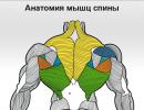
Mimic and chewing muscles:
1 - tendon helmet;
2 - temporal fascia;
3 - temporalis muscle;
4 - occipital-frontal muscle: a) frontal belly, b) occipital belly;
5 - muscle wrinkling the eyebrow;
6 - circular muscle of the eye;
7 - back ear muscle;
8 - nasal muscle: a) alar part, b) transverse part;
9 - muscles of the cheekbones: a) a small zygomatic muscle, b) a large zygomatic muscle;
10 - muscle that lifts the upper lip;
11 - muscle that raises the corner of the mouth;
12 - buccal muscle;
13 - circular muscle of the mouth;
14 - chewing muscle;
15 - muscle lowering the corner of the mouth;
16 - chin muscle;
17 - muscle that lowers the lower lip
3 The work of facial muscles
Scheme of the work of facial muscles

1 - face in a calm state;
2 - a muscle wrinkling the eyebrows;
3 - muscle that reduces yut;
4 - a muscle that lowers the eyebrows;
5 - frontal muscle;
6 - circular muscle of the eye (upper portion);
7 - circular muscle of the eye (lower portion);
8 - circular muscle of the eye (upper and lower portion);
9 - muscle that raises the wing of the nose;
10 - a muscle that expands the wing of the nose;
11 - large and small zygomatic muscles and muscle of laughter;
12 - square muscle of the upper lip;
13 - dog muscle;
14 - circular muscle of the mouth;
15 - triangular muscle.
4. Deep-lying mimic muscles
1. Dog muscle. It is located under the central head of the square muscle. From the infraorbital margin (hard insertion) it extends with fibers to the outer end of the upper lip and partially descends to the outer edge of the lower lip (soft insertion). The canine muscle helps to raise the outer corners of the mouth.
2. Muscle that pulls in and up the upper lip. It has a firm attachment site on the alveolar eminence of the external incisor, and is woven into the tissue of the upper lip with a soft end. Enhances the tightness of the lips.
3. Muscles that lower the wings of the nose. Strain the wings of the nose with a strong retraction of air into the nasal cavity. They have a firm place of attachment - a alveolar elevation of the external incisors, with a soft end attached to the lower outer ends of the wings of the nose.
4. Muscle that besieges the nasal septum. It is attached to the alveolar eminence of the central incisors, and with a soft end to the lower transverse septum of the nose. Precipitates the nasal septum when smelling.
Mimic muscles are the muscles of the face. Their specificity lies in the fact that they are attached at one end to the bones, and at the other - to the skin or other muscles. Each muscle is clothed in fascia - a connective sheath (thin capsule) that all muscles have. What's happened fascia, every housewife can imagine - when cutting meat, we get rid of white films, which, due to their density, worsen its soft texture. In relation to the mimic muscles of the face, in comparison with the muscles of the body, these membranes are so transparent and thin that, from the point of view of classical anatomy, it is believed that the mimic muscles do not have fascia. In any case, the surface of each muscle fiber on the face has a denser structure than its inner part. These connective tissue membranes are woven into the structure of the entire fascial system of the body (through aponeuroses).
It is the contractions of facial muscles that give a variety of expressions to our face, as a result of which the skin of the face shifts and our face takes on one expression or another.
Muscles of the skull
A large percentage of the muscles of the cranial vault is complex in structure supracranial muscle, which covers the main part of the skull and has a rather complex muscle structure. The supracranial muscle is composed of tendon And muscular parts, while the muscular part, in turn, is represented by the whole muscle structure. The tendon part is formed from connective tissue, so it is very strong and virtually indestructible. There is a tendon part in order to stretch the muscle part as much as possible in the areas of its attachment to the bones.
schematically, supracranial muscle can be represented as the following diagram:

The tendon part is very extensive and is otherwise called the tendon helmet or the supracranial aponeurosis. The muscular part consists of three separate muscular bellies:
1) frontal abdomen located under the skin in the forehead. This muscle consists of vertically running bundles that start above the frontal tubercles, and, heading down, are woven into the skin of the forehead at the level of the superciliary arches.
2) occipital abdomen formed by short muscle bundles. These muscle bundles originate in the region of the highest nuchal line, then rise up and are woven into the posterior parts of the tendon helmet. In some sources, the frontal and occipital abdomen are combined into fronto-occipital muscle.

Figure 1. Frontal, occipital abdomen. Tendon helmet.
3) lateral abdomen is located on the lateral surface of the skull and is poorly developed, being a remnant of the ear muscles. It is divided into three small muscles suitable for the auricle in front:
Lateral belly:
- anterior ear muscle moves the auricle forward and upward.
- superior ear muscle shifts the auricle upward, pulls the tendon helmet. A bundle of fibers of the superior auricular muscle, which weaves in a tendon helmet, called temporoparietal muscle . The anterior and superior muscles are covered by the temporal fascia, so their depiction in anatomy textbooks is often difficult to find.
- Posterior auricular muscles A pulls the ear back.

Figure 2. Lateral abdomen: anterior, superior, posterior ear muscles
Muscles of the eye
The muscles of the circumference of the eye consist of three main muscles: eyebrow wrinkling muscleproud muscles and circular muscles of the eye.

Eyebrow wrinkling muscle, starts from the frontal bone above the lacrimal bone, then goes up and attaches to the skin of the eyebrows. The action of the muscle is to reduce the eyebrows to the midline, forming vertical folds in the region of the bridge of the nose.

Figure 3. The muscle that wrinkles the eyebrow.
Muscle of the proud (pyramidal muscle)- originates from the nasal bone on the back of the nose and is attached at the other end to the skin. During contraction of the proud muscles, transverse folds form at the root of the nose.

Figure 4. Muscle of the proud
The circular muscle of the eye is divided into three parts:
- ophthalmic, which starts from the frontal process of the upper jaw, and follows along the upper and lower edges of the orbit, forming a ring consisting of a muscle;
- century- it is a continuation of the circular muscle and is located under the skin of the eyelid; It has two parts - top and bottom. They begin at the medial ligament of the eyelids - the upper and lower edges and go to the lateral corner of the eye, where they attach to the lateral (lateral) ligament of the eyelids.
- tearful- starting from the posterior crest of the lacrimal bone, it is divided into 2 parts. They cover the lacrimal sac in front and behind and are lost among the muscle bundles of the peripheral part. The peripheral part of this part narrows the palpebral fissure, and also smoothes the transverse folds of the skin of the forehead; the inner part closes the palpebral fissure; the lacrimal part expands the lacrimal sac.

Figure 5. Orbital muscle of the eye
Orbicular muscle of the mouth
The circular muscle of the mouth has the appearance of a flat muscle plate, in which two layers are distinguished - superficial and deep. The muscle bundles are very tightly adherent to the skin. Muscle fibers of the deep layer go radially to the center of the mouth.

Figure 6. Orbicularis muscle of the mouth
The superficial layer consists of two arcuate bundles surrounding the border of the lips and repeatedly intertwined with other muscles approaching the oral fissure. That is, in the corners of our mouth, in addition to the fibers of the circular muscles of the lips themselves, the muscle fibers of the triangular and buccal muscles are also woven. This is very important for understanding the biomechanics of aging of the lower part of the face in the section "Spasm of mimic muscles".
The main function of the circular muscle of the mouth is the narrowing of the oral fissure and the extension of the lips.
Muscular system nose
The muscular system of the nose is formed by the following muscles - the nasal muscle, the muscle that lowers the nasal septum, the muscle that raises the upper lip and the wing of the nose.
nasal muscle represented by the transverse and wing parts, which perform different functions.
A) Outer or transverse part, goes around the wing of the nose, expands somewhat and at the midline passes into the tendon, which is connected here with the tendon of the muscle of the same name opposite side.The transverse part narrows the openings of the nostrils. Let's see the picture:
b) The inner, or wing part, attached to the posterior end of the alar cartilage of the nose. The wing part lowers the wing of the nose.>

Figure 7. Transverse and alar parts of the nasal muscle.
Muscle that depresses the nasal septum, most often part of the alar part of the nose. This muscle lowers the nasal septum and lowers down the middle of the upper lip. Its bundles are attached to the cartilaginous part of the nasal septum.

Figure 8. Muscle that depresses the nasal septum.
Muscle that lifts the upper lip and ala of the nose plays a significant role in the formation of nasal folds in team with the nasal muscle and the muscle that lowers the nasal septum. It starts from the upper jaw and is attached to the skin of the wing of the nose and upper lip.

Figure 10. The muscle that lifts the upper lip and wing of the nose.
Cheek muscles
In the area of the cheekbones are the small and large zygomatic muscles, the main function of which is to move the corners of the mouth up and to the sides, forming a smile. Like all facial muscles, both zygomatic muscles have a solid point of upper attachment - the zygomatic bone. At the other end, they are attached to the skin of the corner of the mouth and the circular muscles of the mouth.
Minor zygomatic muscle starts from the buccal surface of the zygomatic bone and is attached to the thickness of the nasolabial fold. By contracting, it raises the corner of the mouth, and changes the shape of the nasolabial fold itself, although this change is not as strong as with contraction of the zygomatic major muscle.

Figure 11. Minor zygomatic muscle
Large zygomatic muscle is the main muscle of laughter. It attaches simultaneously to both the zygomatic bone and the zygomatic arch. The large zygomatic muscle pulls the corner of the mouth outward and upward, greatly deepening the nasolabial fold. Moreover, this muscle is involved in every movement in which a person needs to lift the upper lip and pull it to the side.

Figure 12. Large zygomatic muscle
buccal muscle
The buccal muscle has a quadrangular shape and is the muscular base of our cheeks. It is located symmetrically on both sides of the face. Contracting, the buccal muscle pulls the corners of the mouth back, and presses the lips and cheeks to the teeth. Another name for this muscle - "muscle of the trumpeter", rightly appeared because the muscles of the cheeks affect the compaction and purposefulness of the air stream in musicians playing wind instruments.
The buccal muscle originates from the upper and lower jaws and is woven with another, narrower end into the muscles surrounding the oral fissure. The surface of the buccal muscle from the side of the oral cavity is covered with a thick layer of adipose and connective tissue.

Figure 13. Cheek muscle
Muscle that lowers the corner of the mouth (triangular muscle)
The muscle that lowers the corner of the mouth is located below the corners of the mouth. In shape, it forms a small muscle triangle, which determined its second name - the Triangular muscle. The broad base of the triangular muscle begins at the edge of the lower jaw, and the apex is woven into the circular muscle of the mouth.
The action of this muscle is directly opposite to the action of the zygomatic muscles. If the zygomatic muscles raise the corners of the mouth to create a smile, the triangular muscle lowers the corner of the mouth and the skin of the nasolabial fold. This forms an expression of contempt and displeasure.
Mimic muscles develop from the mesoderm of the second visceral (hyoid) arch. With one end they are attached to the bones of the skull, and with the other end to the skin of the face (see Atl.). These muscles do not have fascia. With their contractions, they displace the skin and determine facial expressions, i.e. expressive facial movements.
Mimic muscles are grouped around the natural openings of the face, one of them covers the roof of the skull. This topography is historically conditioned. In animals - the ancestors of man, the precursors of facial muscles were involved in grasping and holding food, protecting the sense organs from excessive irritations or, conversely, facilitating their perception. In the process of anthropogenesis, these muscles gradually began to express sensations and emotions, especially with the advent of articulate speech among ancient people. Participation in the act of speech determined the differentiation of muscles in the region of the mouth, as well as the eyes. Since the sense of smell in humans is not of leading importance, there was a reduction in the muscles in the nose; the absence of the need to alert the ears led to a reduction in the muscles associated with the auricle.
Mimic muscles include: supracranial (with frontal and occipital bellies); proud muscle; circular muscle of the eye; muscle wrinkling the eyebrow; circular muscle of the mouth; muscle that raises the corner of the mouth; muscle that lowers the corner of the mouth; buccal; muscle that raises the upper lip; zygomatic; laughter muscle; muscle that lowers the lower lip; chin; nose muscles and ear muscles.
Epicranial muscle (t. epicranius) mainly represented by a tendon sprain covering like a helmet (galea), skull roof. Tendon stretch passes into small muscle bellies: behind - occipital (venter occipitales), attached to the upper vyny line; in front - in more developed frontal (venter frontales), woven into the skin of the superciliary arches. If the tendinous helmet is fixed by the occipital bellies, then the contraction of the frontal bellies lays horizontal folds on the forehead and raises the eyebrows (Fig. 1.56). With sufficient development of the abdomens of the supracranial muscle, their contraction sets the scalp in motion.
Muscle of the proud (t. procerus) starts from the back of the nose and attaches to the skin above the bridge of the nose. Contracting, the muscle forms vertical folds here (Fig. 1.56).
Circular muscle of the eye (t. orbicularis oculi) located in the region of the orbit and is divided into three parts: orbital, secular and lacrimal. The orbital part is formed by the most peripheral fibers of the muscle, contracting, they close the eye (Fig. 1.56). The eyelid part consists of fibers laid under the skin of the eyelids; contracting, they close their eyes. The lacrimal part is represented by fibers surrounding the lacrimal sac; contracting, they expand it, which contributes to the outflow of tear fluid into the lacrimal canal.
The muscle wrinkling the eyebrow (i.e. corrugator superecilii), starts from the nasal part of the frontal bone, goes laterally and, piercing the frontal belly of the supracranial muscle, is attached to the skin of the forehead in the region of the superciliary arches. Contracting, the muscle lays horizontal folds on the forehead (Fig. 1.56).
Circular muscle of the mouth (t. orbiculans oris) represents a complex complex of muscle fibers that make up the upper and lower lips. It consists mainly of circular fibers and, contracting, narrows the mouth. Several other facial muscles are woven into the circular muscle of the mouth.
The muscle that raises the corner of the mouth (t. Levator anguh oris), originates from the canine fossa of the upper jawbone. Going down to the corner of the mouth, it attaches to the skin and mucous membranes and is woven into the circular muscle of the mouth in the region of the lower lip.
The muscle that lowers the corner of the mouth (t. depressor anguh oris), originates from the edge of the lower jaw. Converging in its bundles to the corner of the mouth, it attaches to the skin and is woven into the circular muscle of the mouth in the region of the upper lip.
The last two muscles, contracting simultaneously, close the lips.
Cheek muscle (m. buccinator) lies in the thickness of the cheeks. With its upper bundles, it originates from the maxillary bone above its alveolar process, the lower bundles - from the body of the lower jaw below the alveoli, the middle ones - from the maxillary-pterygoid suture - a tendon cord connecting the base of the skull with the lower jaw (see Atl., 30). Heading to the corner of the mouth, the upper bundles of the buccal muscle are woven into the lower lip, the lower bundles into the upper lip, and the middle bundles are distributed in the circular muscle of the mouth. The main value of the buccal muscle is to counteract intraoral pressure. By pressing the cheeks and lips to the teeth, it helps to keep food between the chewing surfaces of the teeth.
Adipose tissue (fat body) accumulates on the buccal muscle, especially in childhood(causes the roundness of children's cheeks).
The muscle that raises the upper lip (t. Levator labii superioris), begins with three heads from the frontal process and the lower orbital edge of the maxillary bone and from the zygomatic bone. The fibers go down and are woven into the skin of the nasolabial fold. Contracting, they deepen this fold, raising and stretching the upper lip and expanding the nostrils (Fig. 1.56).
Large zygomatic muscle (t. zygmaticus maior) goes from the zygomatic bone to the corner of the mouth, which pulls up and to the sides during contraction (Fig. 1.56).
Laughter muscle (t. risorius) inconstant, thin tuft stretches between the corner of the mouth and the skin of the cheek. Contracting, the muscle forms a dimple on the cheek.
Muscle that lowers the lower lip (t. depressor labii interioris) starts from the body of the lower jaw deeper and medially to the muscle that lowers the corner of the mouth; ends in the skin of the lower lip, which, when contracted, pulls down.
Chin muscle (t. mentalis) starts from the holes of the lower incisors, goes down and medially; attached to the skin of the chin. During its contraction, the muscle lifts and wrinkles the skin of the chin, causing the formation of pits on it, presses the lower lip to the upper one (Fig. 1.56).
Nasal muscle (t. nasalis) originates from the holes of the upper canine and external incisor. It distinguishes two bundles: constricting nostrils And expanding their. The first rises to the cartilaginous back of the nose, where it passes into a common tendon with the muscle of the opposite side. The second, attaching to the cartilage and skin of the wing of the nose, pulls the latter down.
Front, top And back muscles of the ear fit the auricle and the cartilaginous part of the external auditory canal. Muscles are rarely developed enough to move the auricle.
Mimic muscles
All facial muscles have the following features: they are devoid of fascia, are woven into the skin, and when they contract, change the position and depth of skin folds. Muscles are under the control of the nervous system and, by contracting, create a certain facial expression that reflects the mental state of a person. Mimic muscles are concentrated near the oral, nasal, orbital and ear openings and are anatomically independent of each other, but in the formation of facial expressions, functional combinations of many muscles are possible that change the position of skin folds and the size of the above facial openings.
Muscles surrounding the mouth
The circular muscle of the mouth (m. orbicularis oris) is a wide circular ribbon starting from the edge of the red border of the lips and reaching the nasal septum at the top, and below the chin-labial groove. This muscle from above, from the side and from below is radially penetrated by numerous muscles, which, contracting, all together stretch the oral fissure. With selective contraction of individual bundles, the shape and position of the lips or the angle of the oral fissure change, which occurs during articulation, eating and chewing.
Function. When contracted, it closes the oral fissure.
The muscle lifting the upper lip and wing of the nose (m. Levator labii superioris alaeque nasi) represents three bundles that start from the wing of the nose, the frontal process of the upper jaw and the infraorbital edge of the zygomatic bone. All three heads are connected near the nasolabial fold, weaving into the skin.
Function. With the reduction of all parts, the nasolabial fold and partially the upper lip rise, which happens with laughter and a smile. In the event that the circular muscle is contracted, it is possible to expand the nasal openings.
The large and small zygomatic muscles (mm. zygomatici major et minor) start from the front surface of the zygomatic bone, go down and medially. The muscle is woven into the middle of the nasolabial fold of the upper lip and the upper part of the angle of the oral fissure.
Function. Pulls the corner of the mouth and raises the nasolabial fold. Participates in the act of laughter.
The muscle that raises the corner of the mouth (m. Levator anguli oris) is the most deep muscle. It begins in the region of the canine fossa of the upper jaw. Attaches to the top of the corner of the mouth.
Function. Raises the corner of the mouth and the upper lip in the region of the incisors.
The muscle of laughter (m. risorius) is represented by a thin and tender bundle that starts from f. parotidis and is woven into the skin of the corner of the mouth.
Function. When contracted, it pulls the corner of the mouth.
The muscle that lowers the lower lip (m. depressor labii inferioris) is a deep muscle. It starts from the base of the lower jaw and is woven into the lower lip.
Function. Lowers lower lip.
The muscle that lowers the corner of the mouth (m. depressor anguli oris) starts from the base of the lower jaw and is attached to the corner of the mouth in the form of a triangular plate.
Function. Lowers the corner of the mouth, smoothing the nasolabial fold and bending the chin-labial fold, which gives the face an expression of sadness and grief or neglect and alienation.
The chin muscle (m. men talis) starts from the alveolar process in the region of the lower incisors and is woven into the skin of the chin.
Function. With bilateral contraction, it lifts the skin of the chin and increases the curvature of the chin-labial fold. The lower lip, resting in this case against the motionless skin of the chin, turns inside out.
The buccal muscle (m. buccinator) limits the vestibule of the mouth. Its muscle bundles are located in a horizontal plane. It starts outside from the alveolar process of the upper jaw, respectively, 1-3 large molars, from raphe pterygomandibularis and crista buccinatoria mandibulae and ends in the circular muscle of the mouth. The muscle is covered with fascia, which is a continuation of the superficial fascia of the neck. At the level of the 2nd large molar of the upper jaw, the duct of the salivary parotid gland perforates the muscle. Outside, between the buccal muscle, skin, angle of the mouth and the coronoid process of the lower jaw, there is a fatty body of the cheek (corpus adiposum buccae), well developed in children.
Function. Pulls the corner of the mouth, flattens both lips, pressing them to the front teeth.
Muscles around the nose
The nasal muscle (m. nasalis) in humans, unlike many mammals, is poorly developed. It starts from the alveolar process of the upper jaw at the level of the second incisor and is divided into three bundles: pars transversa - attaches to the back of the nose, compresses the cartilaginous part of the nose during contraction; pars alaris - attached to the wing of the nose and lowers it; pars depressor septi - attached to the cartilaginous part of the nasal septum, lowers it when contracted.
Muscles surrounding the eye
The circular muscle of the eye (m. orbicularis oculi) is a thin circular plate that has three parts: the orbital, eyelid and lacrimal.
The orbital part (pars orbitalis) starts from the medial ligament of the eyelids, the frontal process of the upper jaw and the nasal part of the frontal bone, then with a wide concentric ribbon is located around the entrance to the orbit. The upper edge of the muscle overlaps the beginning of m. frontalis, lower - the beginning of m. zygomaticus, m. levator labii superioris, descending below the infraorbital foramen.
Function. With the contraction of this part of the muscle, the eyebrow drops, the cheek rises and thus the entrance to the orbit decreases.
The eyelid part (pars palpebralis) is located under the skin of the upper and lower eyelids. It starts from the medial ligament of the eye and goes to the lateral angle, where the bundles mutually intersect.
Function. With the contraction of the secular part of the muscle, the upper eyelid drops by 8-10 mm, and the lower eyelid rises by 2-3 mm. In this case, not only the closing of the palpebral fissure occurs, but also the wetting of the cornea with a tear. The mechanism of this muscle is as follows. With an open palpebral fissure, the curvature of the muscle that surrounds the eyeball is greater than with closed eyelids. Since the beginning and attachment of the muscle are at fixed points, when it contracts, shortening and a decrease in curvature occur, which corresponds to the curvature of the eyeball. As a result, when the muscles contract, the edges of the eyelids tend to take a position in which the curvature of the muscle will be the smallest. The sliding of the eyelids over the eyeball is facilitated by the fact that the conjunctival layers of the eye are moistened with tears.
The lacrimal part (pars lacrimalis) is an underdeveloped muscle. It is located between the upper eyelid and the wall of the orbit. It starts from the crista lacrimalis posterior and, passing behind the lacrimal sac, connects to the posterior edge of the pars palpebralis.
Function. The contraction of the lacrimal part of the muscle exerts pressure on the posterior wall of the lacrimal sac. As a result, the tear is squeezed out of the sac towards the nasolacrimal duct. The action of this muscle is more effective when the palpebral fissure is closed.
The muscle wrinkling the eyebrow (m. corrugator supercilii) is located under the pars orbitalis of the circular muscle of the eye and the final part of m. frontalis. It starts from the nasal part of the frontal bone and the frontal process of the upper jaw, goes to the medial part of the skin of the eyebrow.
Function. When contracting, the muscle not only lowers the medial part of the eyebrow skin, but also brings it closer to the midline. At the same time, oblique or vertical skin folds are formed above the nose.
All the facial muscles described above are innervated by branches of the VII pair of cranial nerves.
Muscles of the outer ear
The muscles of the outer ear in humans are not well developed. This is due to the release of articulatio atlantoaxial mediana, which contributed to an increase in head mobility. The function of moving the auricle towards the sound disappeared.
There are anterior, superior and posterior muscles of the ear (mm. auriculares anterior, superior et posterior). Starting from the fascia, they are attached to the auricle.
innervation: due to the branches of the VII pair of cranial nerves.
Function. When reduced, they cause a slight displacement of the auricle and its tension.
Muscles of the skull
The supracranial muscle (m. epicranius) has two bellies: venter frontalis et occipitalis. Both bellies pass into a wide thin aponeurosis. The frontal abdomen ends in the skin of the eyebrows and, when contracted, raises the eyebrows. The occipital abdomen starts from the linea nuchae superior and, when contracted, stretches the aponeurosis.
The aponeurosis of the supracranial muscle is loosely connected with the bones of the skull and is firmly fused with the skin.
innervation: due to the branches of the VII pair of cranial nerves.
Chewing muscles
Chewing muscle (m. masseter) (Fig. 184), steam room, palpable under the skin in the regio parotideomasseteris. It has a superficial part starting from the zygomatic process of the upper jaw and the zygomatic arch, which is oriented downward and backward and is attached to the angle of the lower jaw, to its tuberosity of the same name. The deep part starts from the zygomatic arch and the deep layer of the temporal fascia and is attached together with the superficial part.
184. Chewing muscles. 1 - m. temporalis; 2 - m. pterygoideus lateralis; 3 - m. pterygoideus medialis; 4 - discus articularis
The chewing muscle develops great strength, as it has a significant moment of rotation.
Function. Raises the lower jaw and is the main muscle in the act of chewing, applying force to the molars.
The temporal muscle (m. temporalis), steam room, has a wide beginning along the entire temporal line of the scales of the temporal bone and a deep sheet of the temporal fascia. Attached to the coronoid process of the lower jaw. The anterior muscle bundles have a vertical position, and the posterior ones are located at an angle of 30 ° to the horizontal line.
Function. The anterior bundles raise the lower jaw and apply force to the incisors and canines. These features of the function can explain the well-developed temporal muscles in predators. The posterior bundles displace the protruding lower jaw backward.
Lateral pterygoid muscle (m. pterygoideus lateralis), steam room, has a triangular shape and is located horizontally. It starts from the infratemporal crest of the greater wing of the sphenoid bone, from the root and outer surface of the lateral plate of the pterygoid process. The muscle bundles are sent back and attached to the pterygoid fossa and the neck of the condylar process. The upper bundles are woven into the joint capsule that covers the articular disc.
Function. With bilateral contraction, the lower jaw is pushed forward. When one muscle contracts, the lower jaw moves in the opposite direction.
The medial pterygoid muscle (m. pterygoideus medialis), steam room, has an orientation of muscle bundles similar to the masticatory muscle. It begins in a fossa bounded by the lateral and medial plates of the pterygoid process. It is attached to the inner surface of the angle of the lower jaw.
Due to m. masseter and m. pterygoideus medialis, a muscular loop is formed, covering the lower jaw in the region of the angle.
Function. Raises the lower jaw, acting mainly on the molars.
The masticatory muscles develop from the mesenchyme of the first gill arch and are innervated by the third branch of the trigeminal nerve.
Fascia of the head
The fasciae of the head are predominantly developed in the masticatory muscles.
Temporal fascia (f. temporalis) is a very dense and durable sheet covering the temporal muscle. It originates from the periosteum along the linea temporalis superior, bifurcates below and is attached by two plates to the inner and outer surfaces of the zygomatic arch.
The fascia of the chewing muscle and the parotid gland (f. parotideomasseterica) covers the muscle of the same name and forms a bed for the parotid salivary gland. The fascia extends upward to the zygomatic process, attaches to the mastoid process behind, passes into the fascia of the sternocleidomastoid muscle, and further from the angle of the lower jaw continues into the fascia of the neck.
The fascia of the buccal muscle (f. buccalis) covers the outer surface of the buccal muscle and the fatty body of the cheek.
Layers of loose fiber of the head
Within the limits corresponding to the cranial vault, loose connective tissue is enclosed between the supracranial muscle (aponeurosis) and the periosteum. This layer of fiber extends from the superciliary arches in front to the upper nuchal line in the back and to the zygomatic arches from the sides.
The periosteum within the frontal, parietal and occipital regions of the head fuses tightly with the bone only near the sutures. In other places between the periosteum and the bones of the skull there is an insignificant layer of loose connective tissue.
In the temporal region, the superficial and deep sheets of the temporal fascia limit the supra-zygomatic interfascial layer, which contains mainly adipose tissue. In the temporal and infratemporal fossae lies the fatty body of the cheek, covered with its own fascial membrane. It is adjacent to the front edges of the temporal and masticatory muscles, continuing to a greater or lesser extent on their outer surface. Part fat body cheeks penetrates between the outer surfaces of both pterygoid muscles on the one hand and the tendon attachment of the temporal muscle, on the other, to the inner surface of the lower jaw branch.
In addition to the fatty body of the cheek, in the temporal and infratemporal fossae there is a large number of loose connective tissue surrounding blood vessels and nerves. This fiber does not have a fascial membrane and, along the course of the vessels and nerves, is introduced between the muscles and into their thickness.
A significant accumulation of loose fiber is located between the lateral pterygoid and temporal muscles. From here, the fiber continues upward, separating the temporalis muscle from the surface of the skull bones. Another accumulation of fiber is located on the inner surface of the lateral pterygoid muscle, which continues into the parapharyngeal. The latter is also closely adjacent to the fiber contained in the capsule of the parotid salivary gland. In the course of the vessels, the loose tissue of the infratemporal fossa passes into the tissue of the pterygopalatine fossa and into the tissue of the capsule of the parotid salivary gland.
Embryogenesis of the muscles of the trunk, head and neck
The development of myotomes occurs in close connection with the solid skeleton of the body, therefore, after the secondary segmentation of sclerotomes and the formation of vertebrae, the deepest myotomes turn into intervertebral muscles. In the anterolateral part of the body, the segmentation of the deep parts of the myotomes, which are transformed into intercostal muscles, is also preserved.
The superficial parts of the myotomes on the dorsal and anterolateral parts of the body lose their segmentation and merge, forming large bookmarks for long and wide superficial muscles torso.
The muscles of the anterior and lateral walls of the abdomen are formed by the fusion of the lower thoracic and upper lumbar myotomes, which on the ventral side have a cranial-caudal orientation. An approximately similar arrangement of muscles is observed in lower animals (see Phylogeny of the muscles of the body). At the 6th week of intrauterine development, the ventral portions of the myotomes fuse into the rectus abdominis muscle. For the rest of their length, they split in the tangential direction, and the deep and superficial parts change the orientation of the muscle fibers. Therefore, the external and internal oblique muscles, as well as transverse muscle abdomen have a different direction of muscle bundles. Such a rearrangement in embryogenesis to some extent repeats the phylogenetic features of the development of the muscles of the lateral wall of the abdomen.
In the initial stages of embryogenesis, the neck is not anatomically isolated and the head is connected directly to the body. If in the region of the body it is possible to distinguish individual segments in the form of sclerotomes and myotomes, then in the region of the head of the four head myotomes, three are involved in the formation of the external muscles of the eyeball. Other muscles of the head develop from the mesenchyme of the head intestine, which has a connection with the cranial nerves. The section "Phylogenesis of the muscles of the head" shows the evolutionary paths of these muscles. In the embryonic period, the mesoderm of the head intestine (pharynx) is divided into sections by gill slits, limited by gill arches (see Development of the skull). Myotomes I and II of the gill arches serve as sources for the formation of the striated muscles of the facial skull (Fig. 185).

The development of facial muscles is associated with the transformation of the mesenchyme of the II branchial arch, innervated by the VII pair of cranial nerves. The rudiment of the mesenchyme of the II branchial arch at the 4-5th week of development extends to the face and neck, forming their subcutaneous muscle, which differentiates into the subcutaneous muscle of the neck and the subcutaneous muscle of the face on the 7th week. The subcutaneous muscle of the face is transformed into mimic muscles, m. stylohyoideus, hind belly - in m. digastricus, m. stapedius, muscles of the soft palate - in m. levator veli palatini and m. uvulae. The muscles of the soft palate are innervated by the X pair of cranial nerves.
The muscles of the larynx and pharynx originate from the mesenchyme of the IV and V gill arches, which are innervated by the IX and X pairs of cranial nerves.
From the ventral part of the occipital myotomes m. trapezius and m. sternocleidomastoideus, while the dorsal ends of the myotomes do not lose their connection with the skull and are innervated by the IXth pair of cranial nerves.
Myotomes on the ventral surface connect to the hyoid bone, give rise to the muscles of the tongue and muscles above and below the hyoid bone.
Phylogenetic development of the muscles of the trunk, neck and head
The complex phylogenetic development of the muscles of the trunk, neck and head is due to the fact that they originate from two rudiments: one part is the own muscles of the trunk, the other is laid in the lateral plates related to the intestinal muscles associated with the mesenchyme of the gill apparatus.
For a more correct representation of the evolution of muscles, we will analyze their rudiments.
The phylogeny of the muscles of the trunk. Lower vertebrates, like the lancelet, have paired muscles located on the sides of the body. Lateral muscles with the help of a horizontal lateral connective tissue septum, they are divided into dorsal and abdominal sections (Fig. 186). Each myomer is separated from the neighboring one by a vertical connective tissue septum (myoseptum); these partitions are located transversely to the body. The vertical septa in the area of the lateral horizontal connective tissue septa are bent and form an angle facing forward. The upper (dorsal) and lower (ventral) ends of the myomeres also curve forward. It turns out a broken line between the myotomes, similar to such a figure, where the angle located on the lateral connective tissue groove is facing forward. In the form of such cones, all the dorsal muscles of the lower vertebrates are built. But already in some fish, differentiation is noted in the position and direction of the myotomes. In the lateral wall of the abdomen, part of the myotomes is located obliquely to the sagittal plane at different depths. This shows that in aquatic animals the first signs of restructuring of myotomes appear, which in terrestrial vertebrates turn into the lateral oblique muscles of the abdomen. In the middle of the abdomen, part of the myotomes is transformed into the rectus abdominis muscle.

186. Superficial abdominal muscles of a newt (according to Maurer). 1 - saphenous muscle; 2- intermuscular septum; 3 - m. obliquus externus superficialis; 4 - m. rectus internus; 5 - m. rectus superficialis
The spinal musculature of terrestrial vertebrates is adapted to the displacement of individual body segments, since the myotomes are thrown over those joints where the movement takes place. In amphibians, the back muscles are built from independent myotomes and resemble the muscles of lower vertebrates. Only in reptiles is the disappearance of the horizontal connective tissue septa and the blurring of the boundary between the dorsal and ventral muscles. Due to increased spinal mobility and chest part of the myotomes merges into more large muscles. This is how mm. interspinales, intertransversales, transversospinales, longissimus, iliocostalis and occipital-vertebral group. This led to the fact that in higher vertebrates, including humans, primary myotomes remained only in the form of short muscles mm. rotatores, connecting successively segments of the body (vertebrae).
The abdominal muscles in lower amphibians still retain division in the form of separate myotomes (Fig. 186), but in the middle there are longitudinal myotomes fused into the rectus abdominis muscle. In the side walls, the myotomes change direction, but do not yet form independent muscle layers. In higher vertebrates, the myoseptae disappear and the myotomes merge with each other into large muscle layers arranged in three layers. These muscle layers in humans are represented by three lateral muscles. Only in the rectus abdominis muscle, in all animals, primary segmentation is preserved in the form of tendon bridges intersectiones tendineae.
IN thoracic region ribs grow along the connective tissue septa between the myotomes, and intercostal muscles are located in the intercostal spaces, representing the continuation of the abdominal myotomes. From the ventral side of marsupials m. pyramidalis reaches the sternum. In mammals, this muscle is preserved as a rudiment.
Phylogeny of the visceral muscles. In lower vertebrates, the anterior intestine is formed by circular fibers covering the entire visceral apparatus in the form of a common constrictor. Starting with cyclostomes and selachians, deep muscle bundles come into contact with visceral arches, which are covered from the outside by lateral muscles perforated by gill openings. IN muscle mass there is a separation of individual muscles: a muscle is attached to the upper part of the I gill arch, which raises the palatine-square cartilage (innervated by the fifth pair of cranial nerves), to lower section- the muscle that leads the lower part of the II branchial arch (innervated by the V pair of cranial nerves), the intermaxillary muscle lying between the branches of the lower jaw (innervated by the V and VII pairs of cranial nerves). From the side of the back behind the gill slits rear end common decompressor is isolated in m. trapezius.
Beginning with amphibians, in connection with the fixation of the upper jaw to the skull, the muscles of the visceral apparatus are transformed. The muscle that lifts the palatine-square cartilage in amphibians is transformed into a muscle that raises the eye, which is reduced in higher animals. From the muscle that leads the lower jaw, chewing muscles are formed.
The intermaxillary muscle in amphibians and reptiles retains its position, and in higher animals it turns into the chin-hyoid muscle. A bundle is separated from the intermaxillary muscle to form the anterior belly of the digastric muscle. All these muscles are innervated by the fifth pair of cranial nerves.
Aquatic animals have muscles that are innervated by the VII cranial nerve. This includes the muscle that lowers the lower jaw, which in mammals turns into the posterior belly of the digastric muscle (innervated by the VII pair of cranial nerves).
In reptiles, the neck constrictor (innervated by the VII cranial nerve) reaches significant development, which in mammals is divided into superficial and deep parts. The muscles of the perioral fissure develop from the deep part, and the rest of the mimic muscles are formed from the superficial part (innervated by the VII pair of cranial nerves).
The muscles associated with the gill apparatus, with the loss of the gill type of breathing, are converted into the muscles of the larynx, pharynx and hyoid. The trapezius muscle loses its connection with the gill arches and switches to shoulder girdle. The sternocleidomastoid muscle splits off from its anterior edge.
In fish and terrestrial vertebrates, the hyoid muscles develop from the abdominal processes of the occipital myotomes, which go around the gill apparatus behind and are located on the ventral side under the visceral apparatus. From the rectus abdominis in amphibians and other more highly organized animals, the muscles of the tongue, the hyoid, and the muscles lying below the hyoid bone develop.
Description of areas
Upper limb belt and upper limb have areas: deltoid (regio deltoidea); acromial (regio acromialis); shoulder, anterior and posterior (regio brac-hii anterior et posterior); ulnar, anterior and posterior (regio cubiti anterior et posterior); cubital fossa (fossa cubitalis); forearm area, anterior and posterior (regio antebrachii anterior et posterior); wrists, anterior and posterior (regio carpi anterior et posterior); metacarpus, palmar and dorsal (regio metacarpi volaris et dorsalis); fingers, anterior and posterior (regio digitorum manus anterior et posterior).
View and buy books on Medvedev ultrasound:
All people have bad and good days, happy and sad events, something happens that angers, offends, upsets or, on the contrary, leads to indescribable delight, causes fun and happiness. At such moments, our face is just a book in which you can read all the feelings.
But why is this happening? What is there in the structure of the face that allows us to be so different, alive, interesting and multifaceted in terms of expression of emotions? Turns out it's merit. different types muscles. It is about them that we will talk in this article.
Number and definition of human muscles
In total, there are about 640 muscles in the human body, each of which performs its own specific function. Muscle anatomy is a collection of complex structural parts.
Muscles (or muscles) are human organs, which are a set of muscle fibers (elongated cells) that have a smooth or transversely striated pattern. They are held together by a loose connective tissue structure. In the human body they form a whole system of skeletal muscles ( striated fabrics) and line many organs and vessels (smooth tissues).

Classification
According to the functions performed, the muscles are divided into the following groups:
- Discharging.
- Leading.
- Arch supports.
- Sphincters.
- Dilators.
- Rotators.
- Flexors.
- Extensors.
- Contrasting.
- Pronators.
There is also a classification of muscles according to their location in the human body. So, allocate:
- trunk muscles (superficial and deep);
- limb muscles;
- head muscles (mimic and chewing).
Form
According to this feature, 7 main muscle groups are distinguished, and each group is localized and functional in a certain part of the human body.
- Fusiform.
- Square.
- Flat.
- Direct.
- Triangular.
- Cirrus.
- Circular.

Muscle Anatomy
Each muscle has roughly the same plan internal structure: on the outside it is covered with epimysium - a special sheath substance produced by connective tissue. From the inside, it is a set of muscle bundles of various orders, which are combined at the expense of endomysium - connective tissue. At the same time, a number of blood vessels and capillaries approach each muscle for an adequate supply of oxygen during work. The veins take away decay products and carbon dioxide. The nerves penetrating the fibers provide conductivity, excitability, and a quick and high-quality response (work).
Muscle cells themselves have several nuclei, since during active work they are able to generate thermal energy due to numerous mitochondria. Muscles owe their ability to contract to special proteins: actin and myosin. It is they who provide this function, causing the contraction of the myofibril - the contractile part of the muscle fiber.
The most important functions of muscle fibers are contractility and excitability, provided by the joint interaction of nerves and protein structures and controlled by the central nervous system(brain and spinal cord).
Muscles of the head
This group includes several main types. The main ones are:
- facial muscles (mimic muscles) - are responsible for facial expressions, external manifestations of emotions;
- chewing - perform the same function.
In addition to them, muscles are distinguished:
- eyeball;
- auditory ossicles;
- language;
- sky;
- pharynx.
The peculiarity of the structure of all the muscles of the head, except for the buccal, is the absence of fascia - a special "bag" in which all the muscles are located and which is attached directly to the bones. Therefore, the vast majority of them are attached to the bones with one side, and the other freely flows directly into the skin, tightly intertwining with it into a single structure.

Mimic muscles of the face: types
The most interesting and clearly showing their work outwardly are just the facial muscles. Due to the function performed, that is, the ability to form human facial expressions, they got their name - facial muscles.
There are enough of them. After all, one has only to remember how bizarre and diverse the expressions of our emotions can be in order to understand that one or two cannot cope with such work alone or together. Therefore, facial muscles act in whole groups, and there are 4 of them in total:
- Forming the vault of the skull.
- Forming the circumference of the mouth.
- Encircling the nose.
- Forming the circle of the eyes.
Let's consider each group in more detail.
Muscles of the calvaria
The facial muscles of the head, which form the cranial vault, are represented by the occipital-frontal, attached to the tendon helmet. The helmet itself is a tendon that conditionally divides the muscle into two parts: the occipital and frontal. The main function that such mimic muscles of the head perform is the formation of transverse skin folds on the forehead of a person.
This group also includes the anterior and posterior ear muscles. Their main action is to enable the auricle to move up, down, forward and backward.
The transverse nuchal muscle is part of the structures of the cranial vault. The main function is the movement of the skin on the back of the head.
Muscles that make up the circumference of the eye
These are the most expressive facial muscles. Their anatomy does not imply the presence of fascia, and the shape of such structures is different.
- The circular muscle completely encircles the eyeball in a circle under the skin. It consists of three main parts: orbital, secular and lacrimal. Action - opening and closing the eyes, control of tear flow, lowering the eyebrows down, smoothing wrinkles on the forehead.
- The facial muscles that wrinkle the eyebrows are attached from the frontal bone to the skin of the eyebrows. Function: the formation of longitudinal folds on the bridge of the nose.
- The muscle of the proud - the name itself speaks of the meaning - forms transverse folds at the base of the nose, giving the face an expression of pride and impregnability.
Such mimic muscles of the face allow people to express their emotions only with their eyes, eyes and skin around them. A lot can be said without words thanks to such features of the structure of the human body.

Muscles that form the circumference of the mouth
No less important are other mimic muscles of the face. The anatomy of this group of muscles is represented by a circular structure surrounding the mouth opening. Several main muscles act here, which are antagonists in relation to each other. This means that some of them expand the oral fissure, and some, on the contrary, narrow it.
- The muscle of the mouth, called the circular. Action: narrowing of the oral fissure and forward movement of the lips.
- Cheek muscles (large and small). Functions: Allow the corner of the mouth to move up, down and sideways.
- Features of the facial muscles of the mouth are that they allow it to move. So, for example, at the base of the upper jaw there is a muscle that allows you to raise the upper lip. Nearby is one that raises the wing of the nose.
- Cheek muscle. Meaning: pulls the corner of the mouth to the side, while contracting from both sides allows you to draw the inner surface of the cheeks to the jaw.
- Laughter muscle. Action: allows the corners of the mouth to stretch laterally.
- Two chin muscles. Features of the mimic muscles of this type are that one of them is unstable and can be reduced. Function: provide movement of the skin of the chin, and also pull the lower lip forward.
- Muscle that lowers the lower lip. Value according to the name.
These are all the main oral facial muscles of the face, the anatomy of which allows a person to smile, talk, express joy and displeasure, and move his mouth.
Muscles around the nose
This group includes only two main muscles:
- nasal muscle, consisting of the inner and outer parts. Action: provide movement of the nostrils and nose;
- muscle that lowers the nasal septum down.
Thus, there are only two facial muscles in the circumference of the nose. Their anatomy is no different from the others discussed above. In general, the listed muscle groups of the eye, mouth, nose and cranial vault are the main components of facial expressions. Thanks to the presence of these muscles, people manage to convey a range of feelings, communicate with each other even without words, and reinforce phrases with the necessary visual expression.
Mimic muscles are very important structures that are also responsible for the formation of wrinkles during the aging process. That is why all the centers involved in plastic surgery and similar procedures invite highly qualified specialists who know the anatomy of the muscles well.

Chewing muscles: varieties
Mimic and chewing muscles are the main components of the face and head. If 17 different structures belong to the first group, then only 4 belong to the second group. However, it is these four chewing muscles that play an important role in human life, as well as in maintaining a beautiful young face oval. Let's take a look at what structures they are.
- The chewing muscle is the strongest muscle trained by a person during a meal. It is located in two parts: deep and superficial. It starts from the zygomatic arch and attaches to the muscles of the lower jaw.
- Temporal - starts from the process of the temporal bone and stretches to the lower jaw.
- Pterygoid lateral - consists of two parts: the upper and lower heads. It starts from the site of the sphenoid bone and ends in the muscles of the lower jaw, forming a complex interlacing with them.
- Pterygoid medial - also located from to the lower jaw.
All these muscles are united by the commonality of their functions, which we will now consider.
Functions
Naturally, since the muscles belong to the chewing group, then their action will be appropriate: ensuring the versatile movement of the jaw:
- Chewing - the lower jaw raises and pushes forward.
- Medial - provides lateral and other movements of the lower jaw.
- Lateral - has similar functions to the medial.
- Temporal - the main assistant in chewing movements. Pulls back the protruding lower jaw, and also allows it to rise up to close with the upper.
In addition, it is the temporal muscle that gives a person a tired, tired and haggard look. If you are in a state of nervous tension, acute feelings and stress for a long time, the body will begin to lose weight, and the face takes on a corresponding haggard expression. This is due to the fact that the temporalis muscle becomes thinner and, being fitted by the skin of the face, visually changes its relief.
Thus, we can conclude that the facial and chewing muscles are the constructors of our face, allowing you to embed any expression, make a variety of movements and change various grimaces. They also allow chewing, which is undoubtedly one of the most important life processes of most living beings, including humans.